This collection harks back to my student days when I spent a lot of time studying cells under a microscope and being dazzled by their beauty, especially when they are artificially stained so that the component parts are not only visible but are in stunning bright colours.
There are 10 pieces in the collection, each about 250mm square and mounted on two long pieces of clear perspex, as if they were on the slides used for looking at them under a microscope. They are mixed media pieces, machine embroidered, made for a module of a City & Guilds qualification.
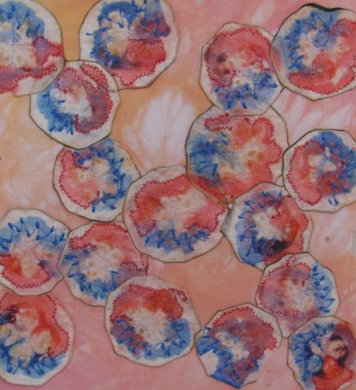
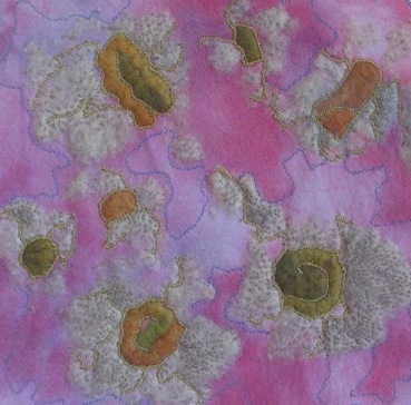
Cells under a microscope:
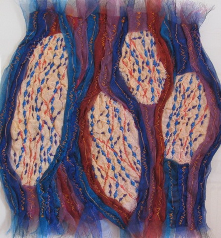
This piece aims to illustrate that view under a microscope. It’s made with very thin slices of courgette which I stained and dried before stitching onto snow dyed fabric.
I was also inspired by a book “The Immortal Life of Henrietta Lacks” by Rebecca Skloot.
It tells the remarkable story of how cancer cells were taken from Henrietta Lacks without her knowledge or consent and how, from these very aggressive cells, scientists were able to reproduce them in an artificial medium for the very first time. That meant that the availability of such cells for research became unlimited. These cells were fundamental in such things as developing the polio vaccine; uncovering the secrets of cancers and viruses, and researching the effects of the atom bomb.
They helped lead to important advances like in vitro fertilization, cloning, and gene mapping; and have been bought and sold by the billions. They were, and still are, called “HeLa” cells, yet Henrietta herself remains virtually unknown, buried in an unmarked grave.
The other pieces in the collection are:
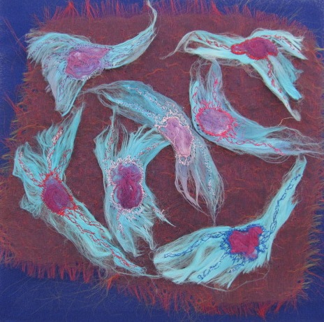
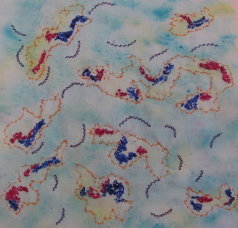
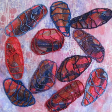
The ground breaking HeLa cells:
Made from dyed silk papers and machine embroidery and mounted on distressed organza.
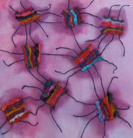
Inspired by Spyrogyra: (simple single celled organisms)
Made from strips and shapes of organza fused together with a soldering iron. The handmade braids were couched on at the end to represent the cell walls.
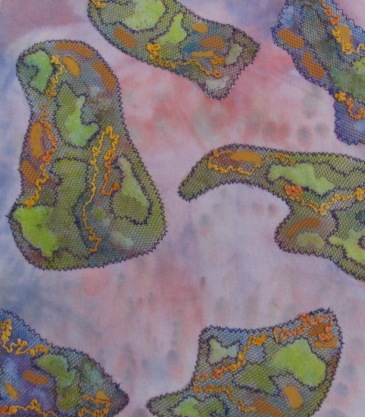
Goli apparatus: the organelles in each cell where proteins are processed.
Made entirely of tissue paper painted with diluted Brusho and scrunched and manipulated with a little oil to make it supple. Beads made from melted organza and lines of machine stitching were added.
Muscle Cells:
The cells were made from embroidered and “crashed” fabric using Solufleece and then machine ebroidered. The long fibres surrounding the cells were made from twisted organzas overstitched by machine.
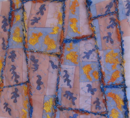
Chromosomes:
Painted Expandaprint and whip stitch were used to create the chromosomes and cell walls respectively, on a background of fabric stippled with wet and dry Brusho.
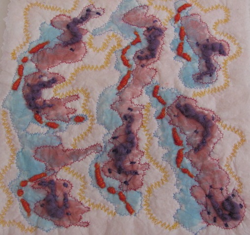
Dispersed cells:
The idea here was to depict cells when they had been separated by centrifugal force.
The cells were constructed with handmade braids and stitched onto wool tops. Then these were mounted onto snow-dyed fabric.
Stained Cells:
This piece was made by cutting back layers of organzas painted fabric and nets with a soldering iron, like reverse applique.
The cells were then mounted on snow-dyed fabric.
Stomata:
Structures on the leaves of plants to allow free transfer of gases for respiration and photosynthesis.
The main components for this piece are the seedheads of Anemone japonica and wool tops trapped under chiffon. These were then mounted on Procion dyed fabric.
Mitochondria:
Vital organelles for respiration.
The walls and crossbars were made by machine stitching onto soluble fabric. They were then stitched onto fabric made from organza scraps, distressed with a heat gun. The whole pieces were then stitched onto a background of white fabric which had been coloured by painting Bondaweb and ironing it on when it was still wet.